Parts of the Eye
Free printable parts of the eye diagram — a labeled cross-section showing the cornea, iris, pupil, lens, retina, optic nerve and more. Download a high-resolution PDF or PNG for Letter paper.
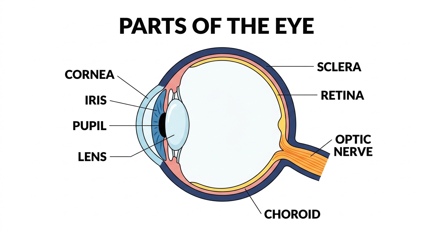
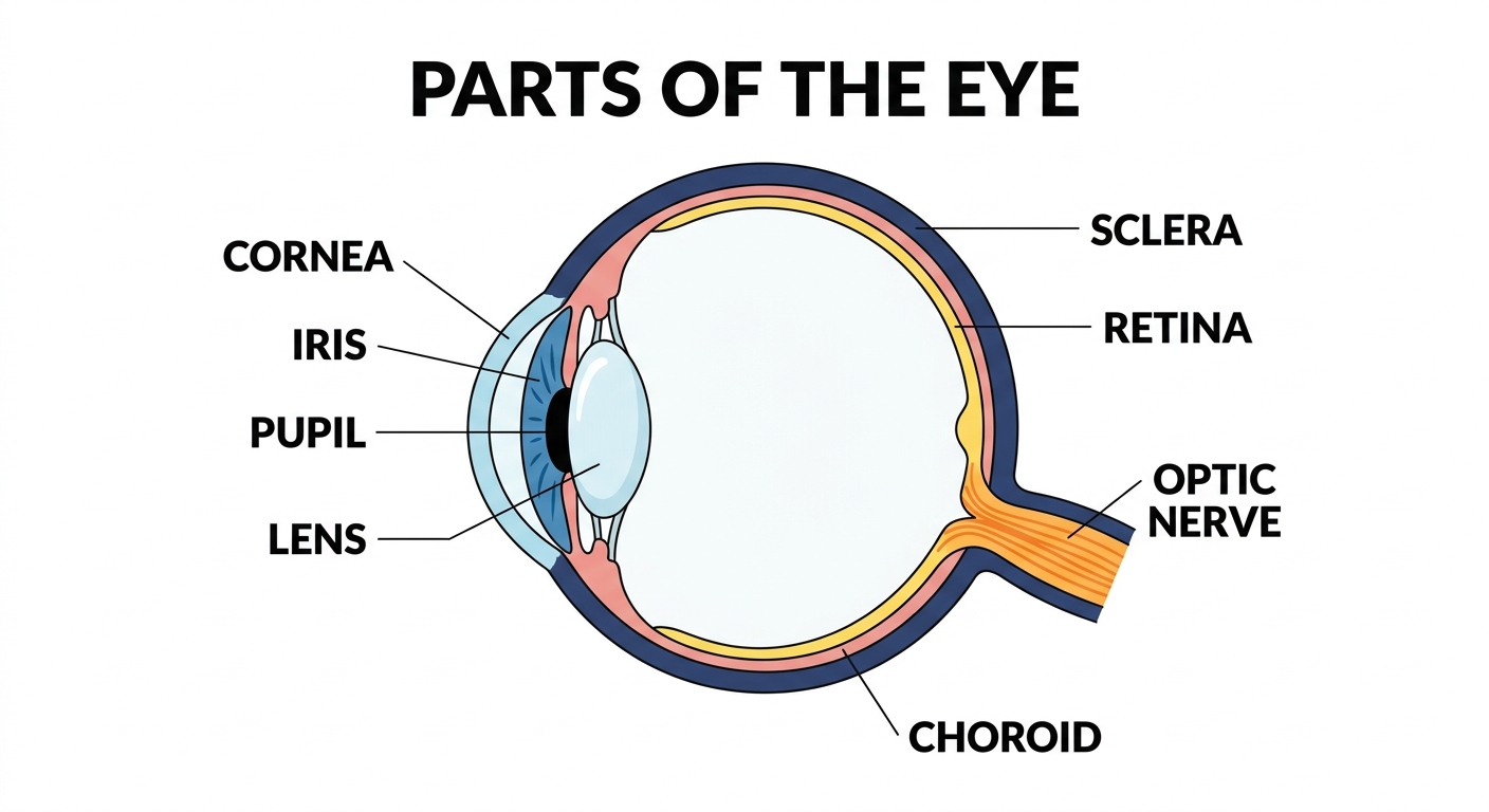
The parts of the human eye
The eye is the organ that lets us see. Light passes through the front of the eye and is focused onto the back, where it is turned into signals sent to the brain. Here is what happens at each labeled part of the diagram:
| Part | What it does | Why it matters |
|---|---|---|
| Cornea | The clear, curved front of the eye that lets light in and helps focus it. | The cornea does about 70% of the eye's focusing work before light even reaches the lens. If it gets scratched or cloudy — from an injury or disease — vision blurs immediately, which is why doctors treat corneal damage as an emergency. |
| Iris | The colored ring of muscle that controls how much light enters the eye. | On a bright beach, the iris squeezes down to protect the retina from being overwhelmed; in a dim room, it opens wide to let in every available photon. No iris control means no ability to adapt — you'd be blinded by sunlight and blind in the dark. |
| Pupil | The black opening in the center of the iris that light passes through. It grows larger in the dark. | It looks like a solid black dot, but it's actually just a hole — you're seeing straight through to the back of the eye. When doctors shine a small flashlight into your eye during a checkup, they're watching whether this opening responds correctly, which tells them a lot about brain and nerve health. |
| Lens | A clear disc behind the pupil that focuses light onto the back of the eye. | Unlike the cornea, the lens can change shape — it flexes to focus on objects near and far, a process called accommodation. Reading glasses exist precisely because the lens stiffens with age and loses that flexibility, making close-up focus harder. |
| Retina | The light-sensitive layer at the back of the eye that turns light into nerve signals. | Packed with roughly 120 million rod cells (for dim-light and motion) and 6 million cone cells (for color and detail), the retina is essentially a living camera sensor. A detached retina — where it peels away from the back of the eye — can cause permanent blindness within days if untreated. |
| Optic nerve | The nerve that carries signals from the retina to the brain. | All those millions of signals from the retina have to travel somewhere, and the optic nerve is the only cable connecting the eye to the brain. There are actually no photoreceptors where it exits the retina, creating your blind spot — a small gap in your vision your brain quietly fills in without you noticing. |
| Sclera | The tough white outer layer that protects the eye and holds its round shape. | This tough, fibrous shell keeps the eye's shape under pressure and anchors the six muscles that move your eye in every direction. Without it, the delicate structures inside would have no rigid frame to sit in — the eye would collapse every time those muscles pulled. |
Companion worksheet
A free fill-in worksheet with an answer key, built from the labels above — students match each part to its description, then answer a couple of short questions. Great for a quick check after using the diagram.
How to print
Choose Download PDF for a ready-to-print file that fits US Letter paper (8.5 × 11") in landscape, or Download PNG for the high-resolution image.
Frequently asked questions
What are the main parts of the eye?
The cornea, iris, pupil, lens, retina, optic nerve and sclera. Light enters through the cornea and pupil, is focused by the lens, and lands on the retina.
What does the pupil do?
The pupil is the opening that lets light into the eye. It grows larger in dim light and smaller in bright light to control how much light comes in.
What paper size should I print this on?
The diagram is sized for standard US Letter paper (8.5 × 11") in landscape orientation. Use "Download PDF" for a print-ready file that fits the page automatically.