Respiratory System Diagram
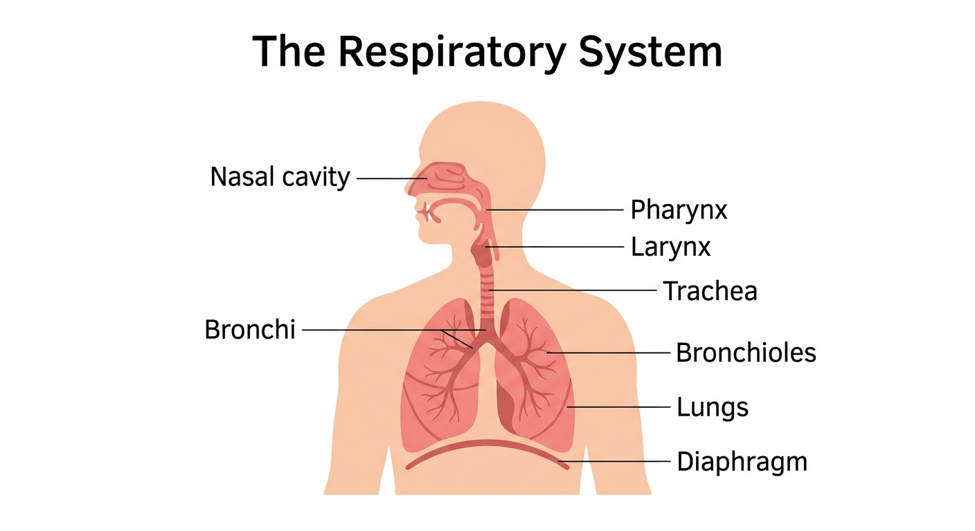
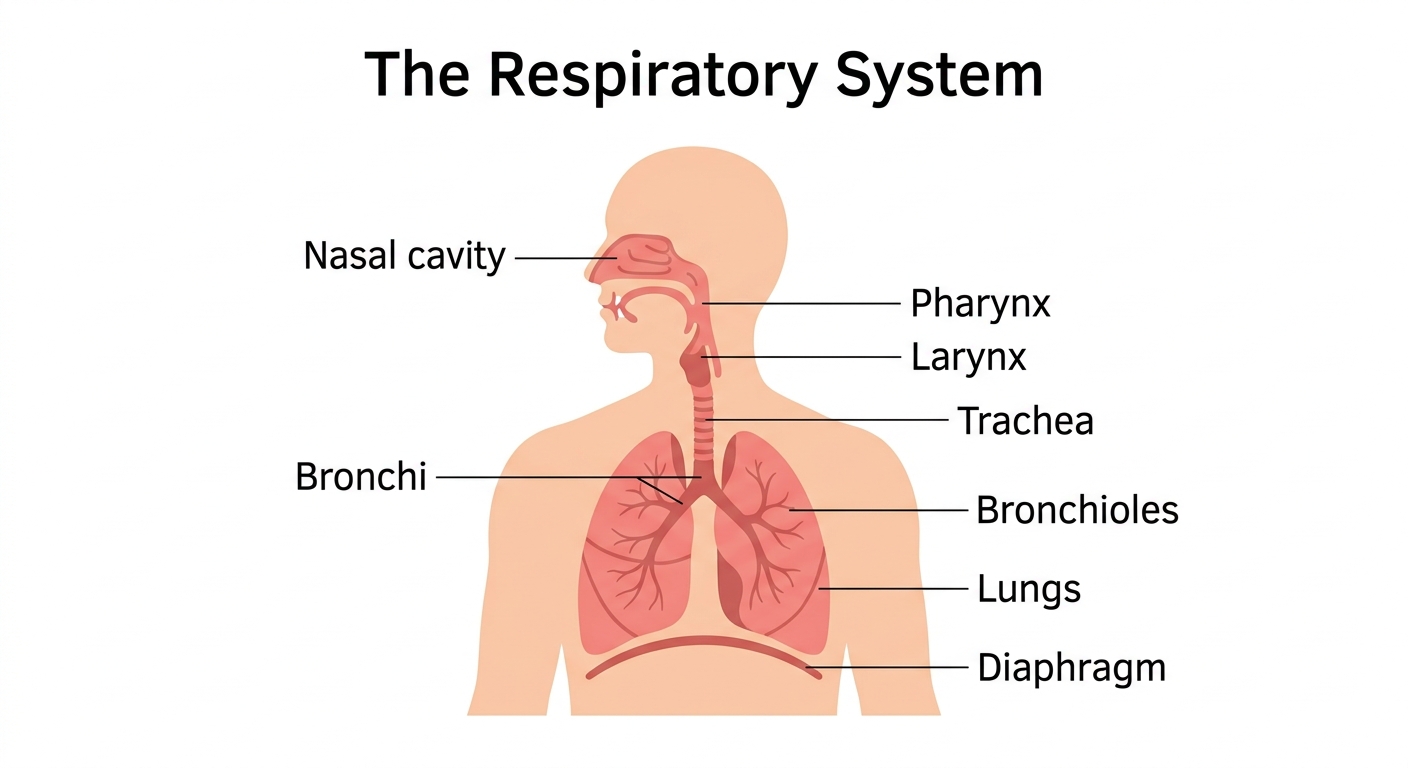
Free printable respiratory system diagram — a labeled illustration of the nasal cavity, trachea, bronchi, lungs, diaphragm and more. Download a high-resolution PDF or PNG for Letter paper.
The parts of the respiratory system
The respiratory system brings oxygen into the body and removes carbon dioxide. Here is what happens at each labeled part of the diagram:
| Part | What it does | Why it matters |
|---|---|---|
| Nasal cavity | Warms, moistens and filters the air you breathe in. | Air straight from outside is often cold, dry, and full of dust — exactly the wrong conditions for your delicate lungs. The nasal cavity fixes all three problems before the air travels an inch further, which is why breathing through your nose during a cold run is actually better for you than breathing through your mouth. |
| Pharynx | The throat — a shared passage for both air and food. | Sharing a passage between food and air is a risky design — food can accidentally enter the airway and cause choking. Your body handles this with careful muscle coordination, but it's why you're told not to talk with your mouth full: switching between the two pathways while swallowing is precisely when things can go wrong. |
| Larynx | The voice box, which also guards the entrance to the airway. | A tiny flap called the epiglottis folds down over the larynx every time you swallow, blocking food from entering the airway — and when you speak, air flowing past the vocal cords inside makes them vibrate, producing sound. Without this dual-purpose guardian, every meal would be a choking hazard and you wouldn't be able to say a word. |
| Trachea | The windpipe that carries air down toward the lungs. | C-shaped rings of cartilage keep the trachea propped open like a garden hose that can't be pinched shut — without them, the tube would collapse every time you inhaled. Doctors can check this passage with a camera, and when it gets blocked by illness or an object, emergency procedures like a tracheotomy restore airflow directly through the neck. |
| Bronchi | Two large tubes that branch from the trachea into each lung. | Splitting the airway into two ensures that each lung gets its own dedicated supply of fresh air, so damage to one side doesn't cut off the other. Conditions like bronchitis inflame the lining of these tubes, making them swell and produce mucus — which is why a chest infection can make every breath feel labored. |
| Bronchioles | Smaller branching tubes deep inside the lungs. | These tiny tubes fan out into hundreds of thousands of branches, multiplying the surface area available for gas exchange far beyond what a simple hollow organ could manage. In asthma, the walls of the bronchioles tighten and squeeze — that's the sensation of an asthma attack, the airways narrowing until airflow slows to a wheeze. |
| Lungs | The main organs of breathing, where oxygen passes into the blood. | Each lung contains roughly 300 million microscopic air sacs called alveoli, and their combined surface area is about the size of a tennis court — all folded inside your chest. That enormous surface is what allows enough oxygen to pass into your bloodstream fast enough to keep every organ in your body running at once. |
| Diaphragm | The dome-shaped muscle below the lungs that powers breathing. | When the diaphragm contracts and flattens, it pulls air into the lungs like a pump; when it relaxes and domes back up, it pushes air out — breathing is almost entirely the work of this single muscle. A hiccup is just the diaphragm contracting in a sudden, uncontrolled spasm, snapping the vocal cords shut and making that unmistakable sound. |
Companion worksheet
A free fill-in worksheet with an answer key, built from the labels above — students match each part to its description, then answer a couple of short questions. Great for a quick check after using the diagram.
How to print
Choose Download PDF for a ready-to-print file that fits US Letter paper (8.5 × 11") in landscape, or Download PNG for the high-resolution image.
Frequently asked questions
What does the respiratory system do?
It takes oxygen from the air into the body and removes carbon dioxide, the waste gas the body makes.
What is the diaphragm?
The diaphragm is a large dome-shaped muscle under the lungs. When it tightens and flattens, air is pulled into the lungs; when it relaxes, air is pushed out.
What paper size should I print this on?
The diagram is sized for standard US Letter paper (8.5 × 11") in landscape orientation. Use "Download PDF" for a print-ready file that fits the page automatically.