Circulatory System Diagram
Free printable circulatory system diagram — a labeled illustration of the heart, arteries (red), veins (blue), and major blood vessels with flow direction arrows. Download a high-resolution PDF or PNG for Letter paper.
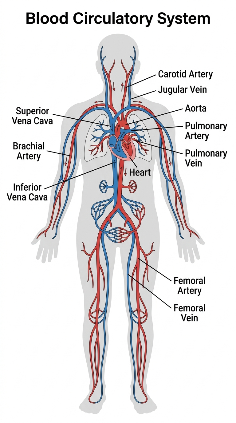

The parts of the circulatory system
The circulatory system pumps blood continuously around the body, delivering oxygen and nutrients to every cell and carrying waste products away. Here is what happens at each labeled part of the diagram:
| Part | What it does | Why it matters |
|---|---|---|
| Heart | The muscular pump at the centre of the circulatory system — it beats about 100,000 times a day. | About the size of your fist and tucked slightly left of centre in your chest, the heart is the most tireless muscle in the body. It contracts and relaxes roughly 100,000 times every day — 35 million times a year — without ever taking a rest. If you live to 80, your heart will have beaten more than 2.5 billion times, pumping enough blood to fill about 200 swimming pools. |
| Aorta | The largest artery in the body, carrying oxygen-rich blood from the heart to the rest of the body. | The aorta leaves the top of the heart and arches over it like a cane before descending through the chest and abdomen. It branches repeatedly into smaller and smaller arteries that reach every tissue in the body. At its widest point the aorta is about as wide as a garden hose — its walls are correspondingly thick to withstand the surge of pressure with every heartbeat. |
| Pulmonary Artery | Carries oxygen-poor blood from the heart to the lungs. | This is the one artery in the body that carries deoxygenated blood — the opposite of every other artery — which is why it is sometimes coloured blue rather than red on diagrams. It leaves the right ventricle of the heart and splits almost immediately into left and right branches heading to each lung, where the blood picks up fresh oxygen and drops off carbon dioxide. |
| Pulmonary Vein | Returns oxygen-rich blood from the lungs back to the heart. | These are the only veins in the body that carry oxygenated blood, and there are four of them — two from each lung — all draining into the left atrium of the heart. From there the freshly oxygenated blood is pumped into the left ventricle, which pushes it out through the aorta to the rest of the body. |
| Superior Vena Cava | Returns oxygen-poor blood from the upper body and head back to the heart. | Formed by the merging of the two large veins draining the arms, head, and neck, the superior vena cava empties into the right atrium — the receiving chamber of the heart. Blood arriving here has already given up most of its oxygen to the tissues of the upper body and is a darker red than the bright-red oxygenated blood leaving the heart. |
| Inferior Vena Cava | Returns oxygen-poor blood from the lower body and legs back to the heart. | The largest vein in the body, the inferior vena cava runs alongside the aorta through the abdomen, collecting blood from the legs, kidneys, and digestive organs before emptying into the right atrium. The digestive organs add a twist — blood draining the intestines first passes through the liver via the portal vein, allowing the liver to process nutrients and remove toxins before the blood continues to the heart. |
| Carotid Artery | Supplies blood to the brain and head. | Two carotid arteries, one on each side of the neck, carry about 70 percent of the blood reaching the brain. You can feel the pulse of the carotid by lightly pressing the side of your neck — this is often where doctors and paramedics check for a pulse in an unconscious person. If blood flow through the carotids is blocked even briefly, unconsciousness can follow within seconds. |
| Femoral Artery | The main blood supply to the leg, running through the thigh. | The femoral artery is one of the largest arteries in the body and the primary blood supply for the entire lower limb. Because it runs close to the surface of the thigh and is large, uncontrolled bleeding from the femoral artery can be fatal within minutes — it is one of the injuries that battlefield medics treat first. Its pulse is also used to assess circulation in the legs during medical examinations. |
Companion worksheet
A free fill-in worksheet with an answer key, built from the labels above — students match each part to its description, then answer a couple of short questions. Great for a quick check after using the diagram.
How to print
Choose Download PDF for a ready-to-print file that fits US Letter paper (8.5 × 11") in portrait, or Download PNG for the high-resolution image.
Frequently asked questions
What does the circulatory system do?
It pumps blood around the body to deliver oxygen and nutrients to every cell, and carries carbon dioxide and other waste products away to be removed.
What is the difference between arteries and veins?
Arteries carry blood away from the heart (usually oxygen-rich, shown in red); veins carry blood back to the heart (usually oxygen-poor, shown in blue).
What paper size should I print this on?
The diagram is sized for standard US Letter paper (8.5 × 11") in portrait orientation. Use "Download PDF" for a print-ready file that fits the page automatically.