Animal Cell Diagram
Free printable animal cell diagram — a labeled animal cell showing the nucleus, mitochondria, ribosomes, Golgi apparatus and other organelles. Download a high-resolution PDF or PNG for Letter paper.
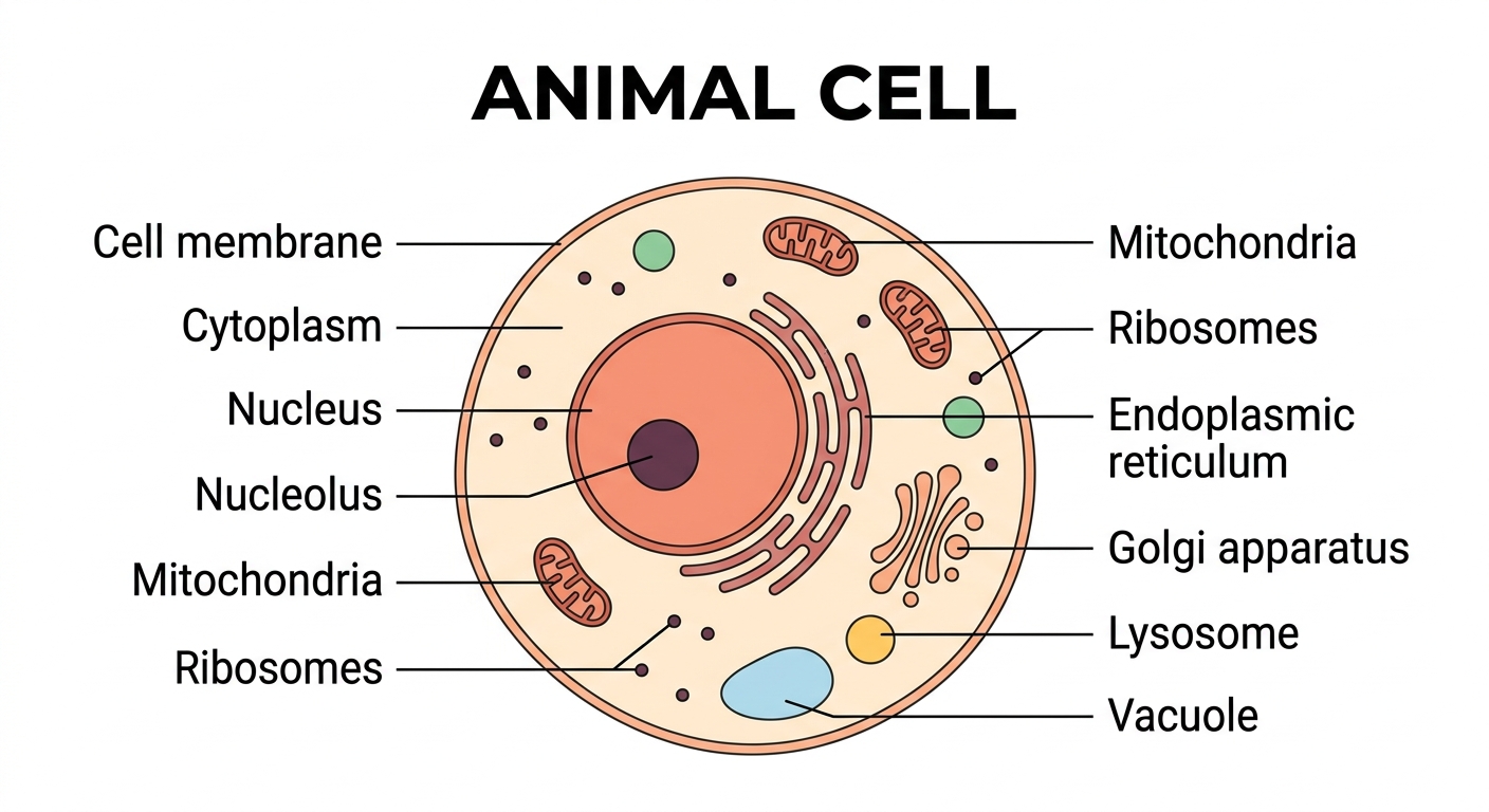
Your body is made of about 37 trillion cells, and every single one of them is running right now. Proteins are the workhorses of life — the enzymes that digest your food, the hormones that carry signals, the materials that build muscle and skin. Most of what a cell does is manufacture the right proteins and get them to the right place. The diagram below shows the key parts that make that happen — and why each one matters.
The parts of an animal cell
An animal cell is the basic building block of animals, including humans. Inside are tiny structures called organelles, each with its own job. Here is what happens at each labeled part of the diagram:
| Organelle | What it does | Why it matters |
|---|---|---|
| Cell membrane | The thin outer layer that holds the cell together and controls what goes in and out. | Without a selective barrier, the cell couldn't maintain the chemical balance it needs to survive — everything would leak in and out uncontrolled. |
| Cytoplasm | The jelly-like fluid that fills the cell and holds the organelles. | It's the medium through which chemicals, proteins, and signals travel between organelles — without it, the cell's parts couldn't communicate. |
| Nucleus | The control center of the cell; it holds the DNA and directs the cell's activities. | When a cell needs to build a protein, it reads instructions from DNA here first. A mutation in the nucleus can disrupt the entire cell's behavior. |
| Nucleolus | A small, dense area inside the nucleus that makes ribosomes. | Ribosomes are the cell's protein-building machines — the nucleolus is the factory that makes those machines before they go to work. |
| Mitochondria | The "powerhouses" that release energy from food for the cell to use. | Every time you move a muscle or think a thought, mitochondria are releasing the energy that makes it possible. Muscle cells, which need lots of energy, are packed with hundreds of them. |
| Ribosomes | Tiny structures that build proteins for the cell. | Proteins run nearly everything in your body — enzymes, hormones, structural materials. Ribosomes are the only place in the cell where proteins are actually built. |
| Endoplasmic reticulum | A folded network of membranes that moves materials around the cell. | Acts as the cell's internal highway, carrying newly-made proteins and fats from where they're produced to where they're needed. |
| Golgi apparatus | Packages and ships proteins and other materials to where they are needed. | The cell's post office: it sorts, labels, and dispatches proteins — some stay inside the cell, others get exported to the bloodstream or neighboring cells. |
| Lysosome | Holds chemicals that break down waste and worn-out parts. | Without lysosomes, damaged proteins and old organelles would pile up inside the cell. They also act as a first defense against bacteria that get inside. |
| Vacuole | A storage sac for water, food and waste. Animal cells have small vacuoles. | While animal cells have small vacuoles, plant cells have one enormous central vacuole — the main reason plants can store so much water and hold their shape. |
Companion worksheet
A free fill-in worksheet with an answer key, built from the labels above — students match each organelle to its description, then answer a couple of short questions. Great for a quick check after using the diagram.
How to print
Choose Download PDF for a ready-to-print file that fits US Letter paper (8.5 × 11") in landscape, or Download PNG for the high-resolution image.
Frequently asked questions
What are the main parts of an animal cell?
The cell membrane, cytoplasm, nucleus, nucleolus, mitochondria, ribosomes, endoplasmic reticulum, Golgi apparatus, lysosomes and vacuoles.
What is the difference between an animal cell and a plant cell?
Plant cells have a rigid cell wall, a large central vacuole and chloroplasts for photosynthesis. Animal cells have none of these — just a flexible cell membrane and small vacuoles.
What paper size should I print this on?
The diagram is sized for standard US Letter paper (8.5 × 11") in landscape orientation. Use "Download PDF" for a print-ready file that fits the page automatically.